
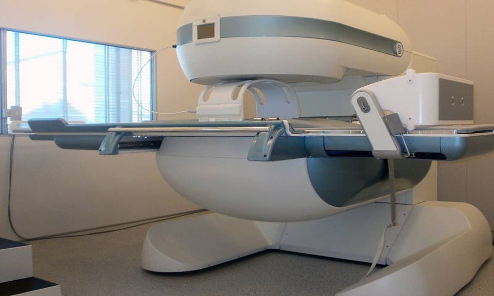
A standard MRI is performed with the patient lying flat, completely still, with the spine and joints decompressed and unloaded. For many conditions, that position is perfectly adequate for diagnosis. For a specific group of conditions, however, the supine position actively works against an accurate diagnosis, because the structures being imaged behave differently once gravity and normal body weight are removed from the equation.
A weight-bearing MRI, performed while the patient sits, stands, or bends, captures the body in the positions where symptoms actually occur. For the conditions below, that difference is not a minor technical detail. It is often the difference between a scan that explains the patient’s pain and one that appears unremarkable despite ongoing symptoms.
1. Lumbar Spinal Stenosis
Spinal stenosis involves narrowing of the spinal canal that compresses the nerves running through it. This narrowing is frequently dynamic, meaning it changes depending on the position of the spine. Many patients with stenosis find that their symptoms worsen significantly when standing or walking and ease when sitting or lying down. A supine MRI captures the spine in its most decompressed state, which can show a canal that appears wider than it actually is during the activities that trigger the patient’s pain. A weight-bearing scan captures the canal under the load that produces the symptoms.
2. Degenerative Spondylolisthesis
Spondylolisthesis is the forward slippage of one vertebra relative to the one below it. The degree of slippage in degenerative spondylolisthesis often increases under axial load, meaning the vertebra shifts further out of alignment when the patient is standing compared to lying down. Grading the severity of spondylolisthesis from a supine scan can understate the true extent of the instability, which has direct implications for treatment planning, particularly when surgical intervention is being considered.
3. Cranio-Cervical Instability
Cranio-cervical instability involves abnormal movement at the junction between the skull and the upper cervical spine. By definition, this is a positional condition: the instability manifests under the load and movement that occurs when the head is upright and unsupported. A supine MRI, with the head resting on a table and the neck supported, removes precisely the conditions under which the instability is most apparent. Weight-bearing MRI in Deerfield allows for upright imaging of the cranio-cervical junction, capturing the anatomy under the gravitational load that produces the patient’s symptoms of headache, dizziness, and neck pain.
4. Herniated Discs That Are Position-Dependent
Not all disc herniations are static. Some discs bulge or herniate only under the compressive load of standing or sitting, and retract partially when the patient lies down and the spine decompresses. A patient with this pattern may have a supine MRI that shows a relatively mild disc bulge, while their actual day-to-day experience involves significant nerve compression symptoms that occur specifically when they are upright. A weight-bearing scan captures the disc in the loaded state where the herniation is most pronounced.
5. Scoliosis and Spinal Curvature Assessment
The degree of spinal curvature in scoliosis changes meaningfully between a supine and a standing position. Gravity acts on the curve when the patient is upright in a way that does not occur when lying flat. For assessing the true severity of a curve, planning treatment, or monitoring progression over time, imaging the spine in the position where the curve is most representative of the patient’s actual posture provides more clinically useful information than a supine measurement alone.
6. Whiplash-Related Ligament Injuries
Whiplash injuries frequently involve ligamentous damage in the cervical spine that does not produce structural changes visible on a standard static MRI. The instability or laxity that results from ligament injury often only becomes apparent when the spine is moved into flexion or extension, positions that a weight-bearing MRI system can capture directly. Patients who have persistent neck pain following a whiplash injury, with a normal-appearing standard MRI, are often good candidates for this type of positional assessment.
7. Chronic Low Back Pain with Normal Standard Imaging
Perhaps the most common scenario where weight-bearing MRI adds value is the patient with chronic low back pain whose standard MRI has come back essentially normal or shows only mild, non-specific degenerative changes that do not seem to explain the severity of their symptoms. This disconnect between imaging findings and reported pain is a recognised challenge in spinal medicine, and patient position during imaging is one of the contributing factors. A weight-bearing scan of the same patient sometimes reveals findings, dynamic stenosis, increased slippage, or position-dependent disc involvement, that were simply not visible in the supine position.
If you have a condition that fits this pattern, symptoms that change with position, or a standard MRI that does not seem to match your experience, it is worth understanding more about conditions an upright MRI can detect that a standard scan may miss entirely. Discussing weight-bearing imaging with your physician is a reasonable next step when your symptoms and your imaging results do not seem to align.